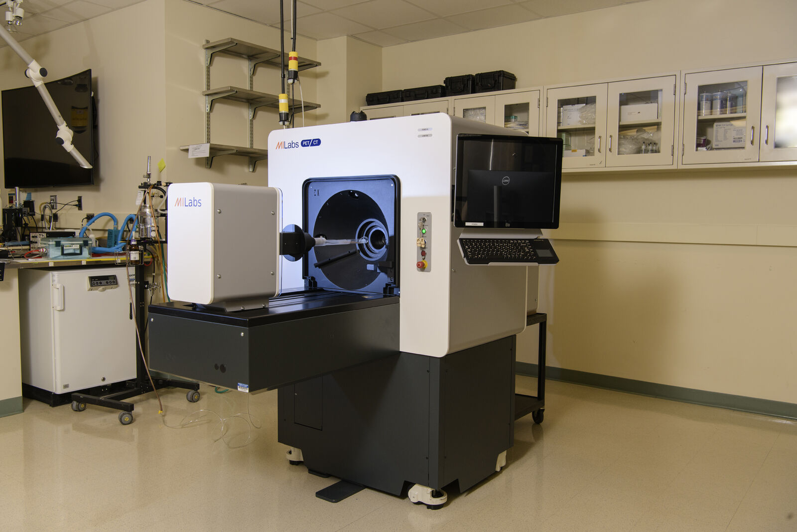
MILabs U-PET7-CT scanner

The central feature of the molecular imaging facility is a hybrid MILabs U-PET7-CT scanner. The scanner is highly adaptable, allowing for various multimodal configurations combining anatomical CT with functional PET imaging modalities.
The central feature of the molecular imaging facility is a hybrid MILabs U-PET7-CT scanner. The scanner is highly adaptable, allowing for various multimodal configurations combining anatomical CT with functional PET imaging modalities.
About the scanner
The system is a hybrid small animal dedicated MILabs U-PET7-CT microPET/CT scanner featuring ultra-high resolution (HE-UHR-M, spatial resolution 0.75 mm, 1.4% sensitivity) and general-purpose high-sensitivity (HE-GP-RM, spatial resolution: 2.4 mm, 3.2% sensitivity) collimators.
It also includes a veterinary monitoring system, enabling rapid evaluation of the in vivo biodistribution and specific uptake of radiotracers within the organ of interest.
The CT component facilitates the acquisition of dynamic (<1 min scan), dual-energy (25-80kV), and high-resolution (<100 um) X-ray CT anatomical images, which can be co-registered with corresponding PET datasets. Controlled by the acquisition workstation, the U-PET7-CT scanner is also equipped with a high-performance server and PMOD, a comprehensive set of user-friendly and powerful tools for image processing, analysis, and quantification.
About the facility
The MILabs U-PET7-CT scanner is housed in a state-of-the-art laboratory space that allows the complete development, synthesis, bioconjugation, and testing of multimodal targeted agents to monitor biological processes within living animals.
The facility encompasses molecular and cell biology resources, bioconjugation, and radiochemistry, including biosafety hoods and radiochemistry hoods with dose calibrators. The suite also contains dedicated areas for sterile surgery and physiological monitoring.
Molecular Imaging Lab contact
BIOMEDICAL IMAGING CENTER
1215 Beckman Institute, 405 N. Mathews Ave., Urbana, Illinois 61801
217-244-0600